I watched the new documentary of David Attenborough “Life in Color”, available on Netflix. I will recommend anything starred by Attenborough, but this one is special for me, so I will leave here the trailer for you:
So, humans have good vision for colors when compared to other mammals. But our color vision is far behind birds’ vision, and a lot of other groups, including some insects. As you know, we can see the rainbow specter, from red to violet. Other organisms may see other colors, including infrared and ultraviolet wavelengths. In this link, you can see some ultraviolet pictures, taken with special cameras, that show a little bit of how much more information than us a bee can get when seeing a flower.
I was 21 years old when I took my first Ishihara test in a neurophysiology class. Ishihara test is a series of numbered plates written in colored dotted patterns and is used to detect color blindness. Funny thing is: I am colorblind, and I discovered it only at that moment. Specifically, I have deuteranomaly. That means that my green cones (special cells that detect green light) do not work properly. I never noticed it before, as well as most of the 6% of XY people. I see green, but not the same way that most of you see. I am also afraid that I cannot really show you any simulation of how I see the world. You may find them on the internet, but they always seem different from the original image for me. So just believe: I see things in a different way. Less shades of green, and some shades of orange-brown-red may be confusing.
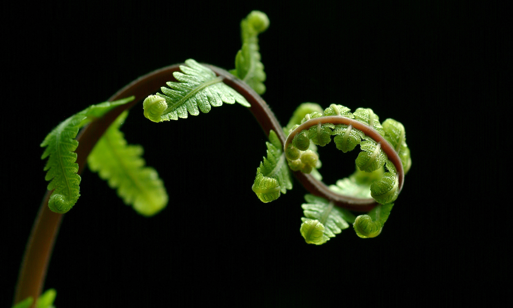
I probably can count in a single hand the number of problems that I had with that before. The worst when I was collecting algae and I could not distinguish the brown ones from red ones (a task I was responsible for as an assistant teacher).
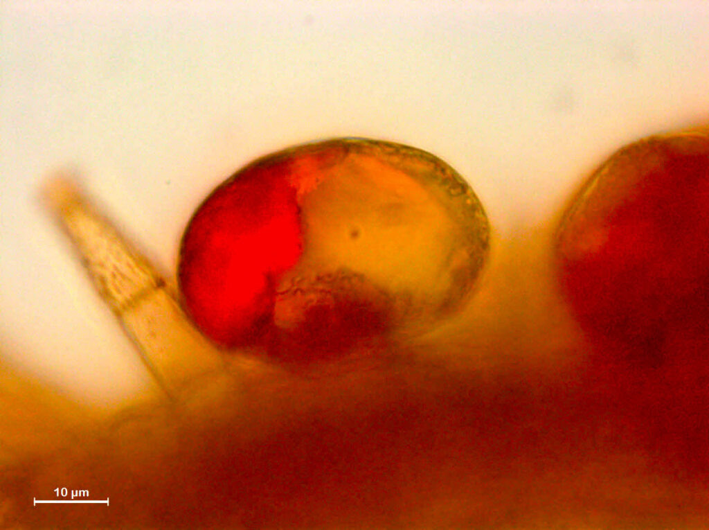
I decided to study structural evolution of plants. And well, I use a lot of Plant Anatomy and microscopy. That means that I slice dead plants in very thin sections, so I can see the organization of tissue and cells in the microscope. We can observe this “fresh” slices, but usually we use some dyes on them. Lot of them are available to detect several compounds: starch, cellulose, lignin (the main component of wood). A lot of colors, a lot of different staining techniques, with beautiful results. One of the most used dyes is Sudan IV, used to detect lipids, and its positive reaction will give you an orange-red color. That is remarkably like brown and green natural colors often found in plant tissues. It means that, when I need to test plant for lipids, I need to borrow my colleague’s eyes. The colors are there, and I trust on them. I respect them, even if I cannot see them. I will leave at the end some good references for several staining techniques in plants 1–5.
When making graphs, schemes, and maps, avoid mixing red/orange and green colors. If you cannot present your pictures in shades of grey or different shapes for dots, try to avoid this specific mix of colors. Doing that, you will make your information accessible to 6% of XY people that see your work. Other types and complete colorblindness are rarer, and these people probably have friends to “borrow” their eyes. If your colorful graph is beautiful, try to upload a “colorblind friendly” supplementary version, if it does not take you much time.
And be aware that sometimes your screen quality does not allow you to see the “true colors”. Have you already been disappointed with printing quality? Sometimes, the problem is not your printer! Monitors with higher values of sRGB are the best and show more trustful colors. Also, the format of images RGB is best for digital images, but CMYK gives better color information for your printer, specifically how much pigment of the four available (cyan, magenta, yellow and black) it should mix. These are different forms of organizing color information in computers. You can easily do this conversion in Photoshop and other image editors, but I can explain better preparation of images for publishing in another post.
Finally, colors are the symbol of LGBTQI+ movements, and this is pride month. I left the closet during the college in the same month that I did that Ishihara test. This is far hard than deal with a deficient color vision, and you need to get over a lot of high school trauma and learn how to trust in people in a (mostly) safer environment. But well, it gets better. And I have my six-colors flag, while I wonder how unique is the way that I see it.
References for staining techniques:
1. Sass, J. E. Botanical Microtechnique. (The Iowa State College Press, 1951).
2. Johansen, D. Plant microtechnique. (McGraw-Hill Book Company Inc., 1940).
3. Ruzin, S. E. Plant Microtechnique and Microscopy. (Oxford University Press, 1999).
4. Kraus, J. E. & Arduin, M. Manual Básico de Métodos em Morfologia Vegetal. Manual Básico de Métodos em Morfologia Vegetal (Editora Universidade Rural (EDUR), 1997).
5. Demarco, D. Histochemistry Analysis of Plant Secretory Structures. in Histochemistry of Single Molecules (eds. Pellicciari, C. & Biggiogera, M.) 313–330 (Springer New York, 2017). doi:10.1007/978-1-4939-6788-9.